How Indian scientists are mapping the brain's last frontier
For more than a century, neuroscientists have studied the human brain much as early cartographers mapped unknown lands: piecing together a vast landscape from scattered
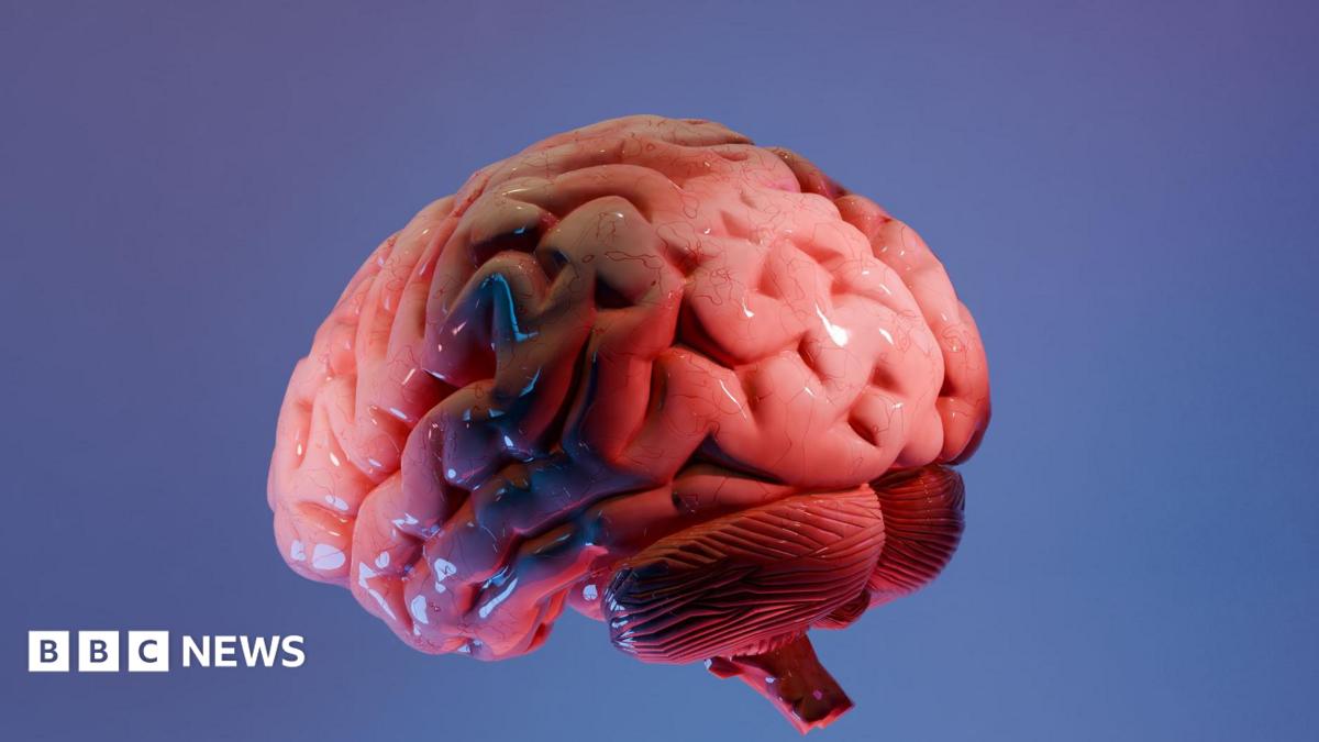
For more than a century, neuroscientists have studied the human brain much as early cartographers mapped unknown lands: piecing together a vast landscape from scattered observations. Even today, pathologists diagnosing disorders such as Alzheimer's disease typically inspect a handful of tissue samples from an organ containing some 86 billion neurons. Much of the landscape remains unseen. That is why scientists at the Sudha Gopalakrishnan Brain Centre (SGBC) at the Indian Institute of Technology, Madras (IIT-M) believe they have taken an important step towards filling one of neuroscience's biggest gaps.
They have produced what they describe as the world's most detailed three-dimensional atlas of the human brainstem at cellular resolution - a digital map that lets scientists travel seamlessly from MRI scans of the whole brain to individual nerve cells. Called Anchor (Atlas of Neurochemical Characterisation of the Human Brainstem with 3D Reconstruction), it combines more than 500 tissue sections from foetal, childhood and adult brains.
Built from high-resolution microscope images rather than costlier molecular techniques, it creates a detailed three-dimensional map of the brainstem, identifying more than 200 clusters of brain cells and nerve pathways. Eight chemical markers help distinguish different cell types, producing one of the clearest pictures yet of this vital, but poorly, understood part of the brain. The brainstem occupies only a sliver of the brain, yet it keeps people alive.
It links the brain to the spinal cord and controls breathing, heartbeat, sleep, wakefulness and movement. Damage to tiny clusters of cells within it can prove catastrophic, but the region's densely packed architecture has long frustrated efforts to map it in detail.
 The BriefWire